Attenuated Total Reflectance (ATR)
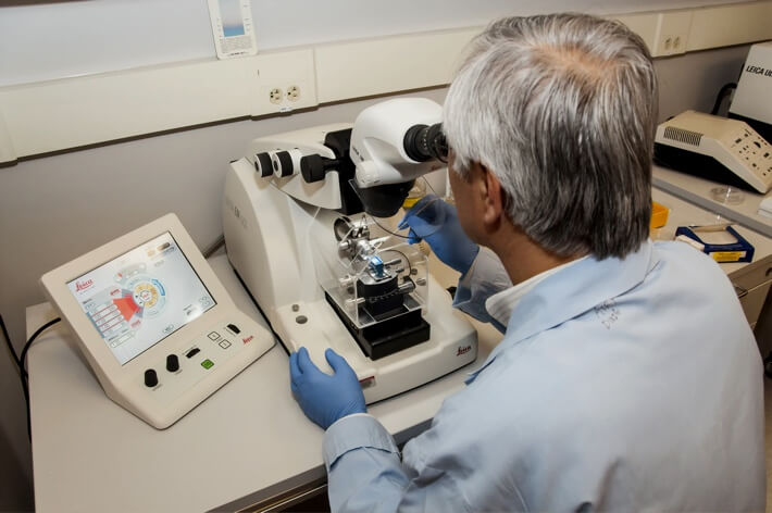
$99 Base price View My Quote RequestTime of Flight Secondary Ion Mass Spectroscopy (ToF-SIMS)
Time of flight secondary ion mass spectroscopy (ToF-SIMS) is a highly surface-specific analytical technique used to qualitatively assess the composition of elements and functional groups within the outermost 1-2 nm of a sample.
Strengths
- Highly surface selective: information depth is 1-2 nm
- Highest trace element / compound sensitivity with detection limit in the ppm and ppb range
- System accommodates insulating and conductive samples
- Non-destructive analysis (outside of depth profiling applications)
Limitations
- Lateral resolution and mass resolution trade-off: optimizing one reduces the other
- Technique is qualitative: meaning it will not precisely report the ratio of elements or compounds, but rather the relative signal intensity from each mass fragment
Technical Specifications:
Learn More:
How ToF-SIMS Works
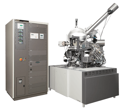
ToF-SIMS is a sensitive and non-destructive (‘static’) variant of a broader class of chemical analysis techniques: secondary-ion mass spectroscopy (SIMS).
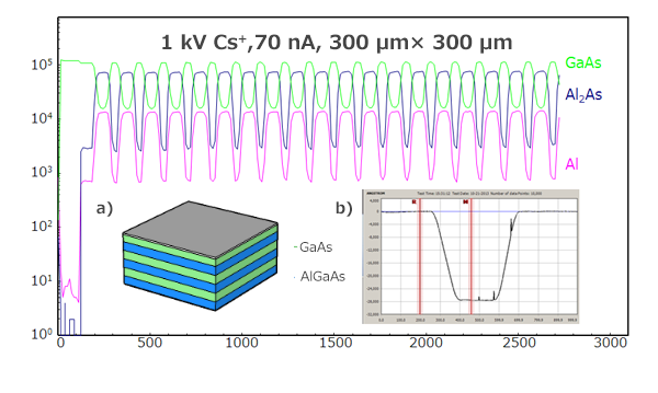
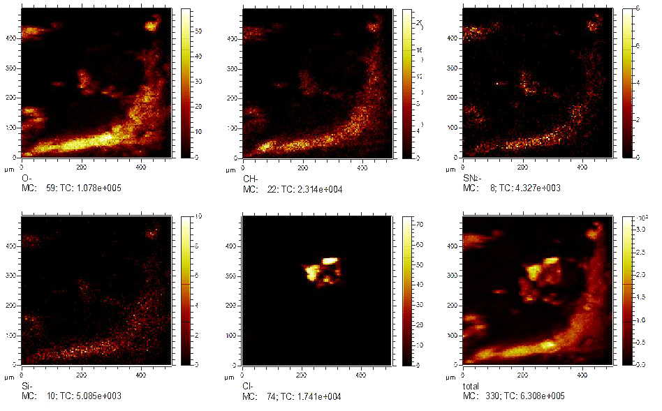
ToF-SIMS instruments use a primary beam of ions scanned across a raster area on a sample to ablate secondary ion fragments from its surface. These secondary ions are then identified according to their mass, generating a spectrum of mass-peaks correlated to the functional groups and elements present in the sample surface.
As the primary beam scans, a total spectrum of ion mass fragments is recorded at each pixel in the raster pattern. This allows for powerful extraction of chemical information from specific regions of interest within maps of each ion species’ relative signal intensity.
+
Add to Comparison
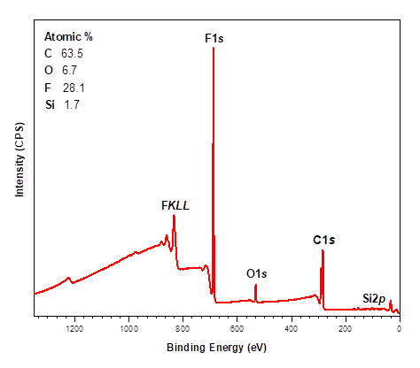
X-ray Photoelectron Spectroscopy (XPS)
In Analytical Chemistry
X-ray photoelectron spectroscopy (XPS) is a highly surface-specific chemical analytical technique used to probe the elemental composition and...
+
Add to Comparison
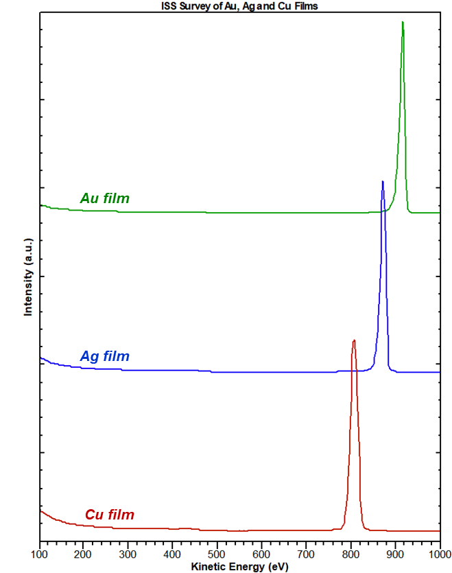
Ion Scattering Spectroscopy (ISS)
In Analytical Chemistry
Ion scattering spectroscopy (ISS) provides quantitative elemental composition information from the very outermost atomic layer of a surface....
+
Add to Comparison
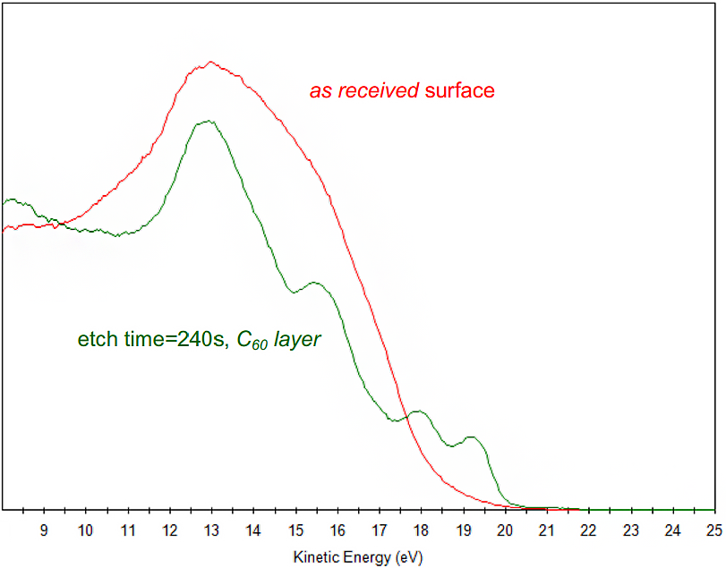
Ultraviolet Photoelectron Spectroscopy (UPS)
In Material Testing
UPS is often performed in conjunction with X-ray Photoelectron Spectroscopy (XPS), a powerful surface chemical characterization technique. Unique...
✕
Comparison link sent successfully
✕
Please use valid email address
✕
You need to have at least 2 techniques to compare
✕
You can select maximum 5 techniques
