Attenuated Total Reflectance (ATR)
$99 Base price View My Quote RequestFIB-SEM: 3D Reconstruction by Tomography

FIB-SEM Tomography produces 3D reconstructed volumes from serial sample images collected using 3D Auto Slice and View TM. It allows internal / subsurface reconstruction of morphological and chemical information on a micro- to nanoscale.
Technical Specifications:
Learn More:
How Works
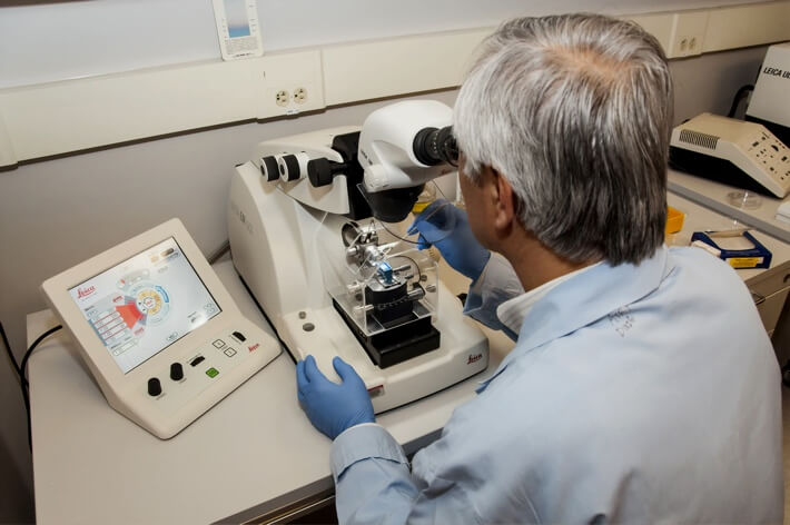
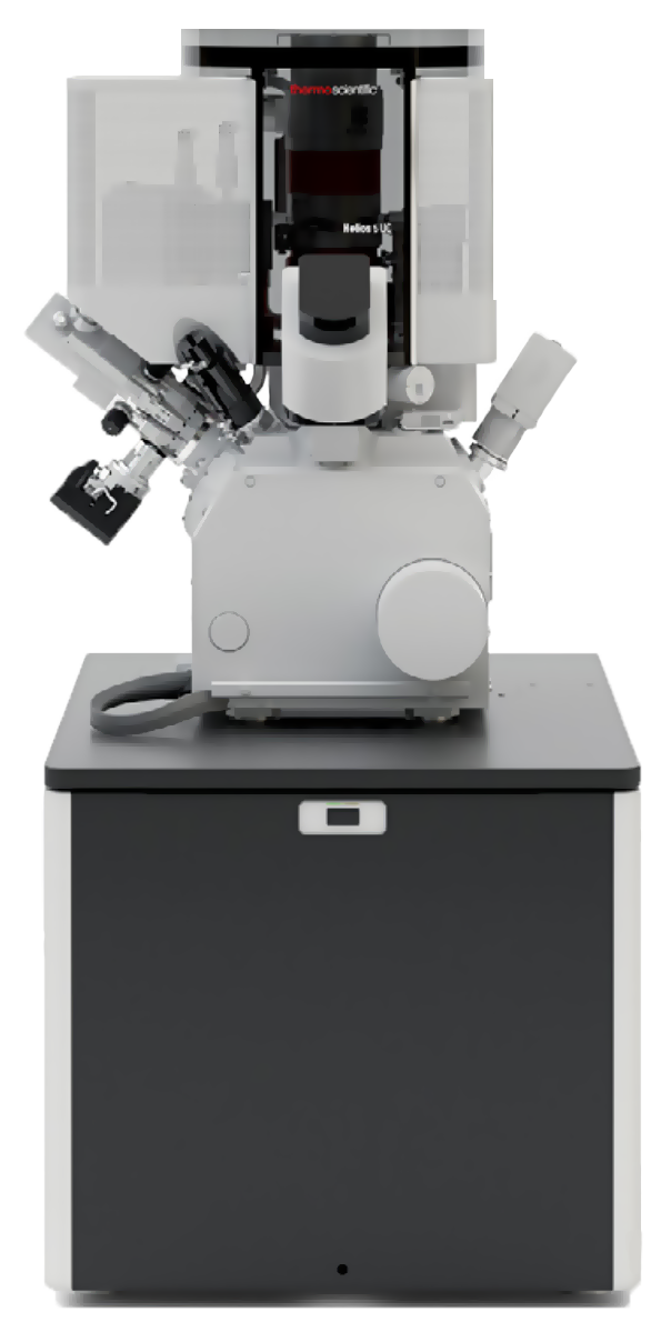
FIB-SEM tomography is performed using automated Auto Slice and View TM software on Thermo Fisher Scientific DualBeam microscopes. This software facilitates serial ion-beam milling of thin layers from a cross-sectional face of a sample, capturing electron micrographs between each cut into the face*. Once the desired region of the sample has been milled and imaged, the software reconstructs a 3D model by stitching the collected 2-dimensional cross-sections into a composite volume. Once the 3D reconstruction is produced, it can be manipulated and analyzed to make dimensional measurements of internal structures and features of interest.
By combining FIB-SEM tomography with EDS or EBSD, analysts can investigate elemental distributions and crystallinity throughout a 3D reconstructed volume. This can be particularly useful in the analysis of alloys, porous materials, or integrated circuits and microprocessors.
* FIB-SEM tomography can cause higher-than-normal charge buildup in certain nonconductive samples. To mitigate this, it may be necessary to sputter coat the sample with a conductive metallic layer. Furthermore, depending on the shape and material of the sample, embedding the sample in resin may improve imaging resolution by fixing its position in a rigid mount.
✕
Comparison link sent successfully
✕
Please use valid email address
✕
You need to have at least 2 techniques to compare
✕
You can select maximum 5 techniques
