Attenuated Total Reflectance (ATR)
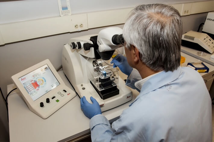
$99 Base price View My Quote RequestElectron Probe Microanalysis (EPMA)
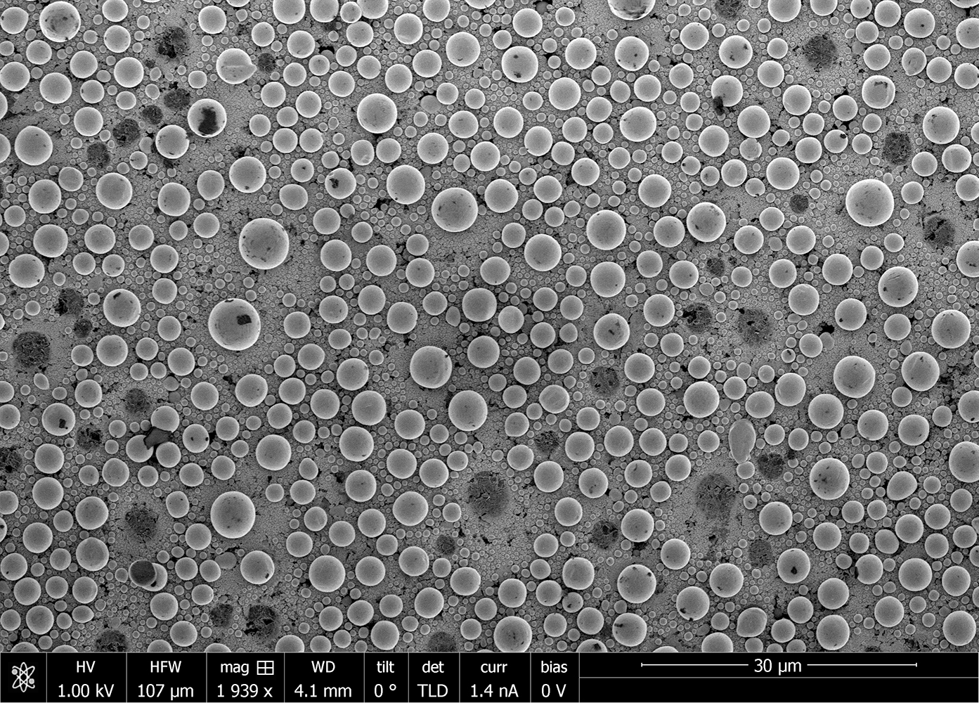
Electron probe microanlysis (EPMA) is a non-destructive technique used for high-sensitivity, quantitative determination of the elemental composition of a material.
Strengths
- Quantitative elemental analysis down to within ~10 to 100 ppm sensitivity
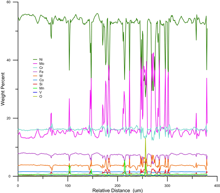
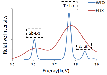
- Simultaneous data collection of up to 5 element signals
- Highest lateral resolution among elemental microanalysis techniques
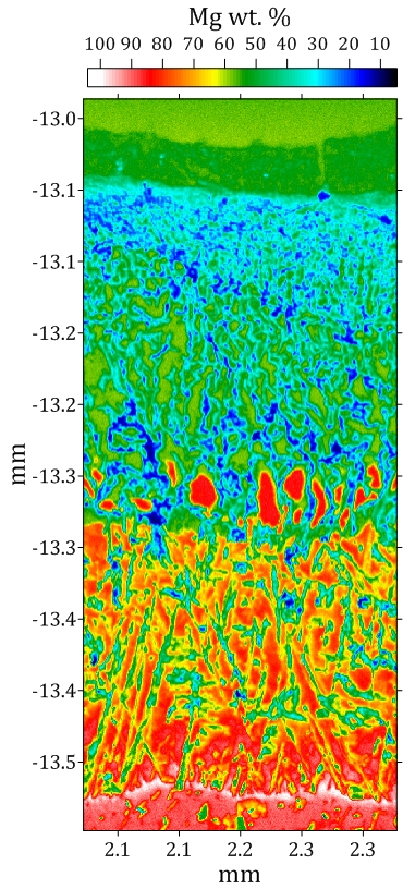
- Elemental mapping enabled
Limitations
- Measurements are time-intensive compared to energy dispersive x-ray spectroscopy (EDS or EDX)
- For quantitative results, elements must be calibrated against standards
- Light elements (Be through N) can be difficult to measure accurately
- Spatial resolution limited to ~ 1 µm
Technical Specifications:
Learn More:
Sample Requirements
- Solid phase
- Must be vacuum stable
- Analyzed surface must be as smooth as possible for best result; polishing sometimes required
- Must be conductive or able to be coated with a conductive material
- Lateral Dimension Range: 1 mm to 1.5 inches
- Vertical Dimension Range: 500 µm to 7/8 inch
How EPMA Works
This technique focuses an energetic beam of electrons onto the sample surface, thereby stimulating x-ray fluorescence. The wavelength of the x-ray is characteristic of the fluorescing element. The strength of that x-ray signal relates then to the abundance of its element within the excited volume, the depth the element is within the sample, and the effects of other elements in the sample.
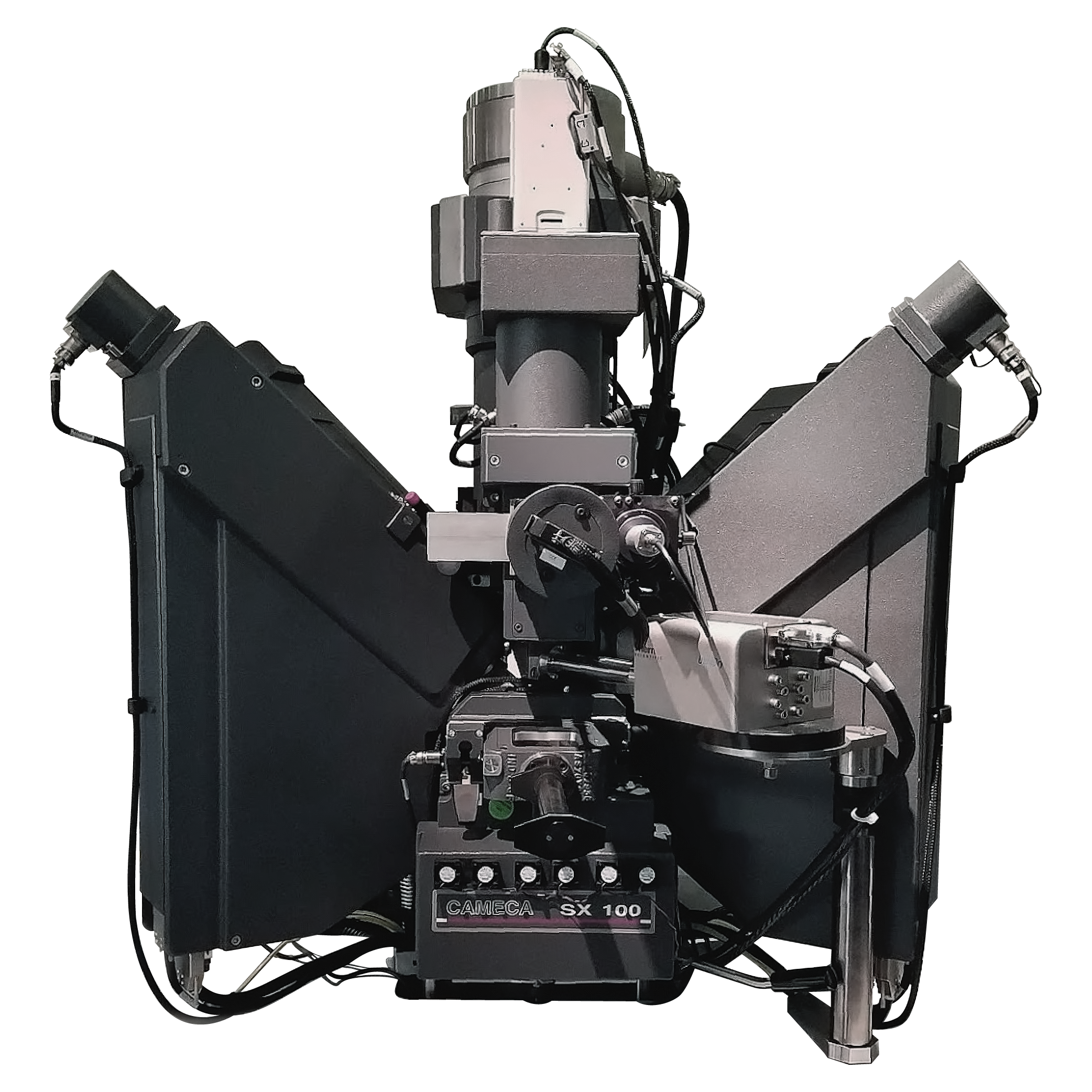
Similarly to Wavelength Dispersive X-ray Fluorescence Spectroscopy (WDXRF), EPMA systems use wavelength-dispersive spectroscopy (WDS) detectors – which act like diffraction gratings – calibrated to detect various x-ray wavelengths. By using wavelength-analyzers, EPMA achieves high resolution separation of emission lines, with low background, allowing for quantitative sensitivity to 10 ppm with appropriate use of reference standards.
Additionally, EPMA systems – like Scanning Electron Microscopes – can be used to generate 2D element maps by measuring fluorescence at multiple points in a raster-scan.
The EPMA system utilized by Covalent also contains an energy dispersive x-ray spectroscopy (EDS) detector that can quickly obtain entire XRF spectra from points on the sample, albeit with much lower spectral resolution and sensitivity. This allows for rapid identification of the elements present to expedite quantitative analysis of their respective concentrations.
+
Add to Comparison
Scanning Electron Microscopy (SEM)
In Microscopy & Imaging
Scanning electron microscopy (SEM) is a surface imaging technique capable of achieving nm resolution on topographical features. Additionally,...
+
Add to Comparison
Wavelength Dispersive X-ray Fluorescence Spectroscopy (WDXRF)
In Analytical Chemistry
Wavelength dispersive x-ray fluorescence spectroscopy (WDXRF) is a non-contact, non-destructive technique used to measure elemental composition, elemental concentration...
✕
Comparison link sent successfully
✕
Please use valid email address
✕
You need to have at least 2 techniques to compare
✕
You can select maximum 5 techniques
