Attenuated Total Reflectance (ATR)
$99 Base price View My Quote RequestAuger Electron Spectroscopy (AES)
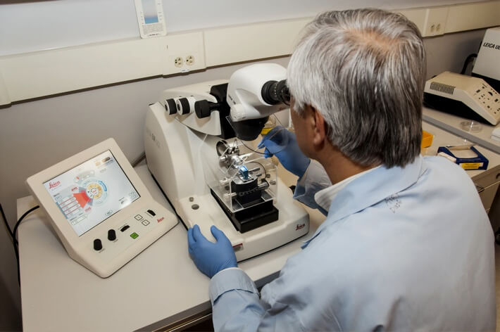
Auger electron spectroscopy (AES) is a surface-sensitive analytical technique with high lateral resolution. It is used to quantify and map the elemental composition of the outermost 2-10 nm of a material.
Strengths
- Very high lateral resolution (as small as 5 nm)
- Provides quantitative elemental composition and chemical state identification
- Acute surface selectivity (typical information depth is ~ 3 to 10 nm)
- Excellent for elemental mapping
Limitations
- Reproducibility can vary
- Charging affects different portions of the spectra by different amounts, which can make analysis difficult
- Analysis is destructive
Technical Specifications:
Learn More:
Instruments Used for AES
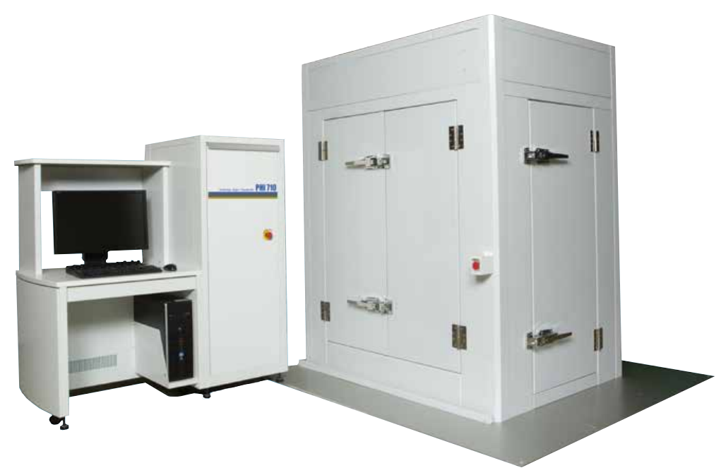
PHI 710 Scanning Auger Nanoprobe
The PHI-710 Auger nanoprobe is a unique AES instrument designed and optimized for high-performance auger spectroscopy applications. It provides superior Auger imaging, spatial resolution, sensitivity, and high spectral energy resolution to address the most demanding AES applications.
How AES Works
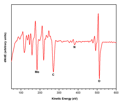
This technique utilizes high-energy electron beam as an excitation source resulting in the emission of Auger electrons, whose kinetic energies are characteristic of the elements and chemical states present in the surface of a sample. The emitted auger electrons are picked up by a specialized detector, which scans over an energy range and analyzes the amount of auger electrons at each kinetic energy value. The resulting spectrum allows analysts to quantitatively determine the elemental composition of the surface. The source electron beam can also be finely focused to diameters as small as ~5 nm to create secondary electron and Auger images which are used not only for compositional and topographic analysis but also to locate features of interests. In conjunction with ion beam sputtering, depth profiling can also be done on samples to provide composition as a function of depth as well as layer thicknesses.

+
Add to Comparison
X-ray Photoelectron Spectroscopy (XPS)
In Analytical Chemistry
X-ray photoelectron spectroscopy (XPS) is a highly surface-specific chemical analytical technique used to probe the elemental composition and...
+
Add to Comparison
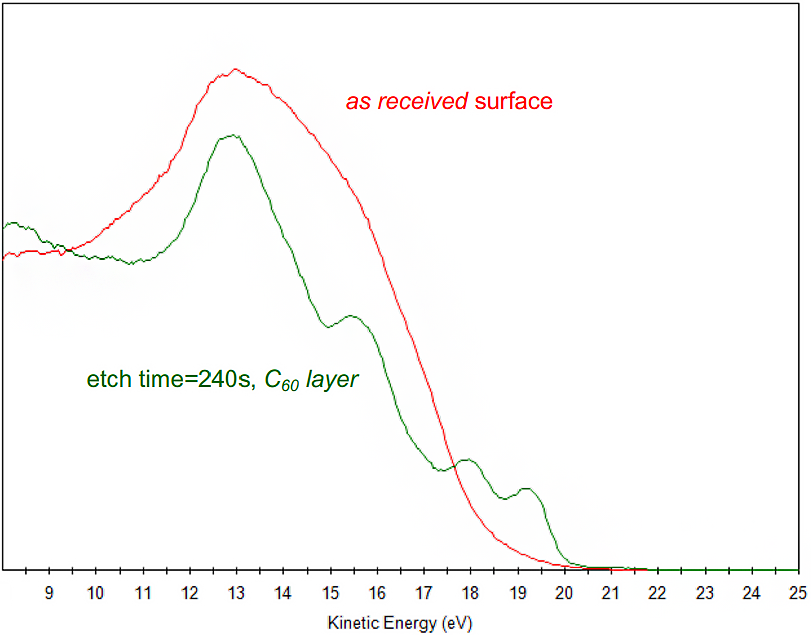
Ultraviolet Photoelectron Spectroscopy (UPS)
In Material Testing
UPS is often performed in conjunction with X-ray Photoelectron Spectroscopy (XPS), a powerful surface chemical characterization technique. Unique...
+
Add to Comparison
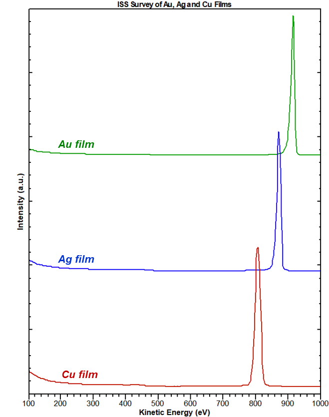
Ion Scattering Spectroscopy (ISS)
In Analytical Chemistry
Ion scattering spectroscopy (ISS) provides quantitative elemental composition information from the very outermost atomic layer of a surface....
+
Add to Comparison
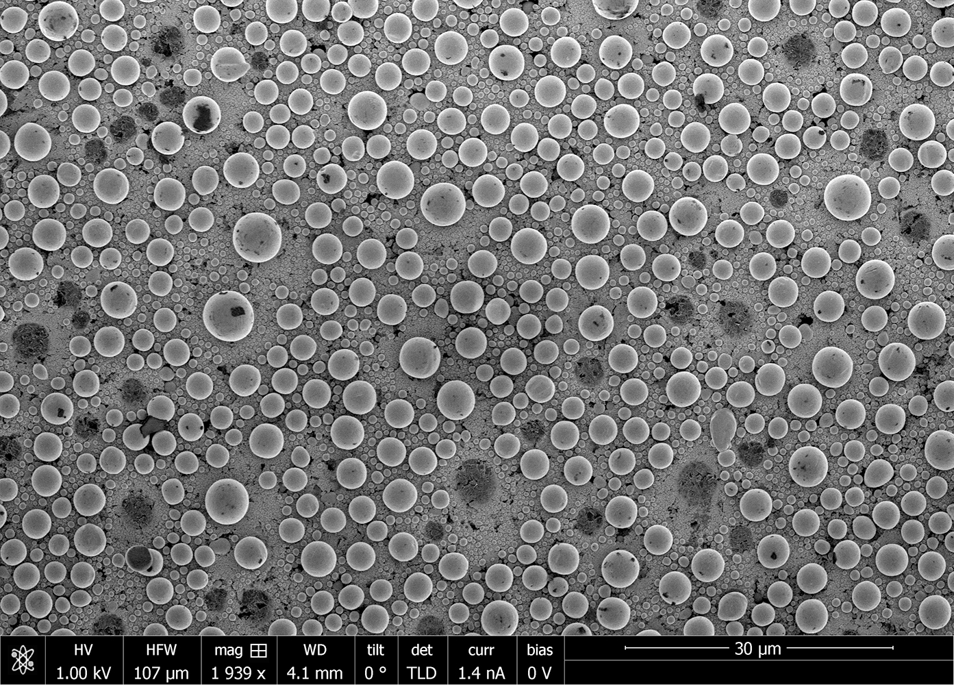
Scanning Electron Microscopy (SEM)
In Microscopy & Imaging
Scanning electron microscopy (SEM) is a surface imaging technique capable of achieving nm resolution on topographical features. Additionally,...
✕
Comparison link sent successfully
✕
Please use valid email address
✕
You need to have at least 2 techniques to compare
✕
You can select maximum 5 techniques
